Introduction
Brain scans are essential tools in modern medicine, providing critical insights into the structure and function of the brain. These imaging techniques help healthcare professionals diagnose various neurological conditions, assess brain injuries, and plan treatment strategies.
Understanding the different types of brain scans and their applications is crucial for both patients and healthcare providers. This article explores the primary types of brain scans, their mechanisms, and their uses in clinical practice.
As we delve into the world of brain scans, we will uncover how these imaging techniques contribute to our understanding of brain health and the management of neurological disorders.
Stay sharp and foster your brain development with our Trivia game generator or Fun Facts game for you mental exercises and mental sharpness!
Overview of Brain Scans
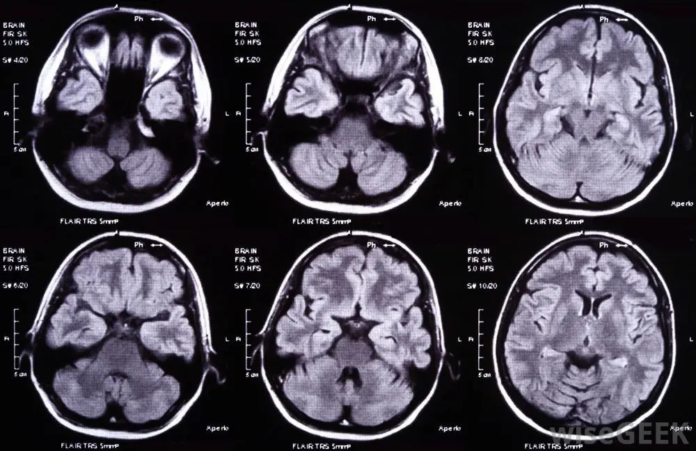
Brain scans can be broadly categorized into two main types: structural imaging and functional imaging. Each type serves a unique purpose and provides different information about the brain.
Structural Imaging
Structural imaging techniques focus on visualizing the anatomical structures of the brain. They provide detailed images that help identify abnormalities such as tumors, lesions, or structural changes.
1. Computed Tomography (CT) Scan
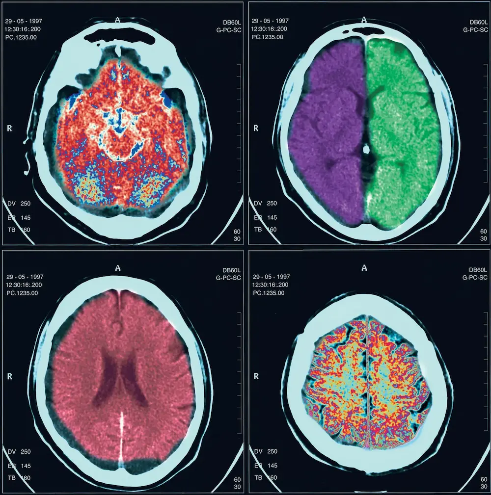
Computed tomography (CT) scans utilize X-ray technology to create cross-sectional images of the brain. The patient lies on a table while the CT scanner rotates around their head, capturing images from multiple angles.
- Uses: CT scans are commonly used to assess head injuries, detect bleeding, and identify tumors or structural abnormalities.
- Advantages: CT scans are quick and can be performed in emergency situations, making them ideal for trauma cases.
2. Magnetic Resonance Imaging (MRI)
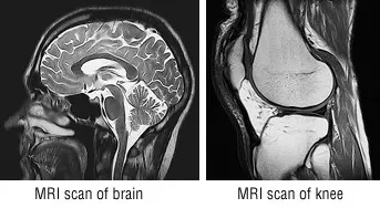
Magnetic resonance imaging (MRI) uses powerful magnets and radio waves to produce high-resolution images of brain structures without ionizing radiation. This technique provides detailed images of soft tissues, making it particularly useful for assessing brain abnormalities.
- Uses: MRI scans are employed to diagnose conditions such as brain tumors, multiple sclerosis, and stroke.
- Advantages: MRI offers superior contrast resolution compared to CT, allowing for better visualization of soft tissue structures.
Functional Imaging
Functional imaging techniques assess brain activity and metabolic processes. These scans provide insights into how different regions of the brain function during various tasks.
3. Positron Emission Tomography (PET) Scan
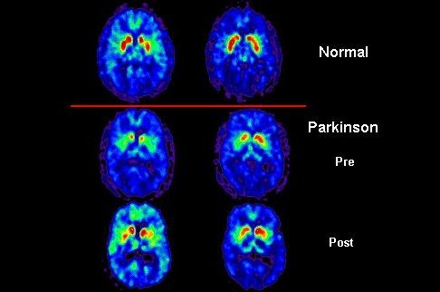
Positron emission tomography (PET) scans involve injecting a radioactive tracer into the bloodstream. The tracer accumulates in areas of high metabolic activity, allowing for visualization of brain function.
- Uses: PET scans are used to evaluate conditions such as Alzheimer’s disease, seizures, and brain tumors.
- Advantages: PET scans provide information about brain metabolism and can help identify areas of abnormal activity.
4. Functional Magnetic Resonance Imaging (fMRI)
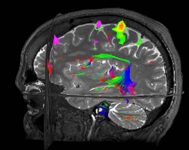
Functional MRI (fMRI) measures changes in blood flow and oxygen levels in the brain, reflecting neural activity. This technique allows researchers to observe brain function in real-time.
- Uses: fMRI is commonly used in research settings to study brain activity during cognitive tasks and in clinical settings to assess brain function before surgery.
- Advantages: fMRI provides high spatial resolution and does not require radioactive tracers, making it a safe option for patients.
5. Electroencephalography (EEG)
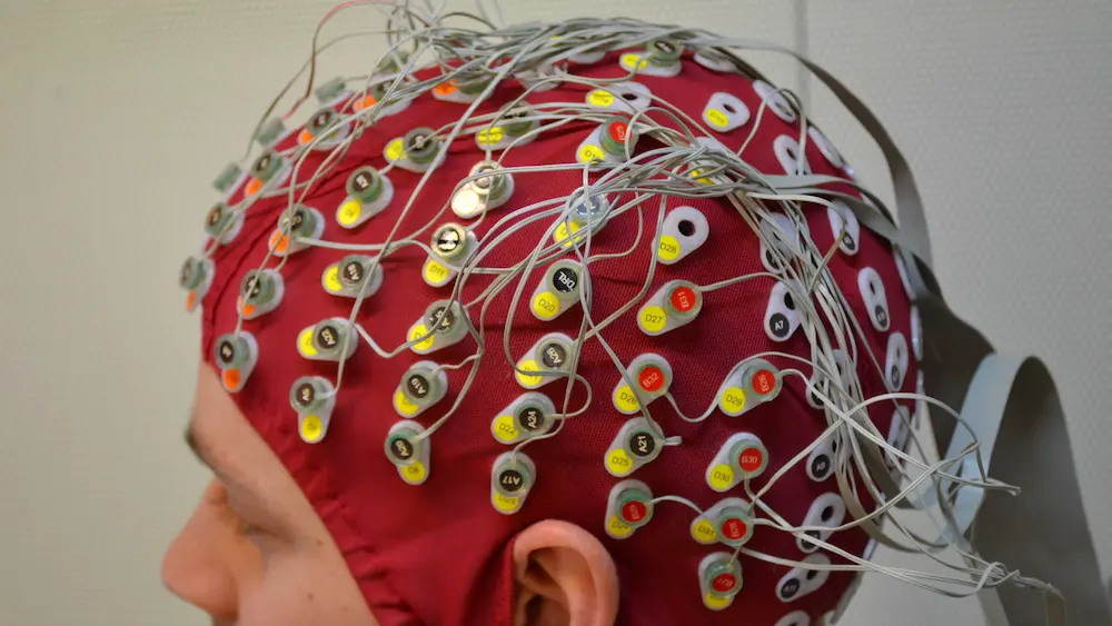
Electroencephalography (EEG) measures electrical activity in the brain using electrodes placed on the scalp. This technique captures brain wave patterns and can help diagnose various neurological conditions.
- Uses: EEG is primarily used to diagnose epilepsy, sleep disorders, and other conditions affecting brain activity.
- Advantages: EEG provides real-time data on brain activity and is non-invasive.
Choosing the Right Brain Scan
The choice of brain scan depends on the specific clinical question and the patient’s condition. Healthcare providers consider factors such as the type of symptoms, the urgency of the situation, and the information needed for diagnosis or treatment planning.
| Brain Scan Type | Primary Uses | Advantages |
|---|---|---|
| CT Scan | Head injuries, bleeding, tumors | Quick, ideal for emergencies |
| MRI | Tumors, stroke, multiple sclerosis | High-resolution images of soft tissue |
| PET Scan | Alzheimer’s, seizures, brain tumors | Measures metabolic activity |
| fMRI | Brain function during tasks, pre-surgical mapping | High spatial resolution, non-invasive |
| EEG | Epilepsy, sleep disorders | Real-time monitoring of brain activity |
Detailed Examination of Each Brain Scan Type
Computed Tomography (CT) Scan
How It Works
CT scans use X-rays to take multiple images of the brain from different angles. A computer then processes these images to create cross-sectional views (slices) of the brain, allowing for detailed examination of its structure.
Clinical Applications
CT scans are particularly useful in emergency settings. They can quickly identify:
- Traumatic Injuries: Detecting fractures, bleeding, or swelling in the brain following an accident.
- Stroke: Differentiating between ischemic strokes (caused by a blockage) and hemorrhagic strokes (caused by bleeding).
- Tumors: Identifying the presence, size, and location of brain tumors.
Limitations
While CT scans are invaluable in emergencies, they do have limitations:
- Radiation Exposure: CT scans expose patients to ionizing radiation, which may pose risks, especially with repeated scans.
- Soft Tissue Visualization: CT scans are less effective than MRI in visualizing soft tissues and certain brain structures.
Magnetic Resonance Imaging (MRI)
How It Works
MRI uses powerful magnets and radio waves to generate detailed images of the brain. It detects changes in the magnetic properties of hydrogen atoms in the body, producing high-resolution images of brain structures.
Clinical Applications
MRI scans are utilized for a variety of conditions, including:
- Brain Tumors: Providing detailed images to assess tumor size, type, and location.
- Multiple Sclerosis: Identifying lesions in the brain and spinal cord associated with MS.
- Neurodegenerative Disorders: Assessing changes in brain structure related to conditions like Alzheimer’s disease.
Limitations
While MRI is highly effective, it also has some drawbacks:
- Cost and Availability: MRI machines are expensive and may not be available in all medical facilities.
- Time-Consuming: MRI scans typically take longer than CT scans, which can be a disadvantage in emergency situations.
- Claustrophobia: Some patients may experience anxiety or discomfort in the enclosed MRI machine.
Positron Emission Tomography (PET) Scan
How It Works
PET scans involve the injection of a radioactive tracer, which emits positrons as it decays. These positrons collide with electrons in the body, producing gamma rays that are detected by the PET scanner to create images of metabolic activity in the brain.
Clinical Applications
PET scans are particularly useful in scanning mental illness and conditions such as:
- Alzheimer’s Disease: Identifying changes in brain metabolism associated with the disease.
- Epilepsy: Localizing areas of the brain responsible for seizures.
- Cancer: Assessing the spread of tumors and monitoring treatment response.
Limitations
Despite their advantages, PET scans have limitations:
- Radiation Exposure: The use of radioactive tracers exposes patients to radiation, although the levels are generally considered safe.
- Cost: PET scans are more expensive than other imaging techniques, which may limit accessibility.
Functional Magnetic Resonance Imaging (fMRI)
How It Works
fMRI measures brain activity by detecting changes in blood flow and oxygenation levels. When a brain region is active, it consumes more oxygen, leading to increased blood flow to that area. fMRI captures these changes in real-time.
Clinical Applications
fMRI is widely used in research and clinical settings:
- Cognitive Neuroscience: Studying brain activity during specific tasks, such as problem-solving or memory recall.
- Pre-Surgical Mapping: Identifying critical brain areas before surgery to minimize the risk of damage to essential functions.
Limitations
While fMRI is a powerful tool, it has some limitations:
- Sensitivity to Motion: Patient movement can affect the quality of images, making it essential for patients to remain still during the scan.
- Complexity of Data Interpretation: Analyzing fMRI data requires specialized knowledge and expertise.
Electroencephalography (EEG)
How It Works
EEG measures electrical activity in the brain by placing electrodes on the scalp. It captures brain wave patterns, providing insights into brain function and activity.
Clinical Applications
EEG is primarily used for:
- Epilepsy Diagnosis: Identifying seizure activity and determining the type of seizures.
- Sleep Studies: Analyzing sleep patterns and diagnosing sleep disorders.
- Brain Function Monitoring: Assessing brain activity during surgeries or in patients with altered consciousness.
Limitations
EEG also has its limitations:
- Spatial Resolution: EEG provides excellent temporal resolution but poor spatial resolution compared to other imaging techniques.
- Interpretation Challenges: Analyzing EEG data can be complex and requires trained professionals.
The Future of Brain Imaging Technologies
Advancements in Imaging Techniques
The field of brain imaging is continuously evolving, with ongoing research aimed at improving existing technologies and developing new methods. Some promising advancements include:
- Hybrid Imaging: Combining different imaging modalities, such as PET/MRI, to provide complementary information about brain structure and function.
- Higher Resolution Imaging: Developing techniques that allow for higher resolution images, enabling better visualization of small structures and abnormalities.
- Artificial Intelligence: Utilizing AI algorithms to analyze imaging data more efficiently and accurately, assisting in diagnosis and treatment planning.
Implications for Research and Clinical Practice
Advancements in brain imaging technologies have significant implications for both research and clinical practice:
- Enhanced Understanding of Brain Disorders: Improved imaging techniques can lead to a better understanding of the underlying mechanisms of various neurological and psychiatric disorders.
- Personalized Medicine: Advanced imaging may enable more tailored treatment approaches based on individual brain characteristics and responses to therapies.
- Early Detection: Enhanced imaging capabilities could facilitate the early detection of brain disorders, allowing for timely intervention and improved outcomes.
Conclusion
Understanding the various types of brain scans is essential for both patients and healthcare providers. Each imaging technique offers unique insights into brain structure and function, aiding in the diagnosis and treatment of neurological conditions.
Leveraging these advanced imaging technologies, healthcare professionals can make informed decisions and provide optimal care for their patients. As brain imaging technology continues to evolve, it will undoubtedly enhance our understanding of the brain and improve outcomes for individuals with neurological disorders.
The integration of advanced imaging techniques into clinical practice will pave the way for more effective diagnosis, treatment, and management of brain health.
Frequently Asked Questions (FAQs)
What are brain scans used for?
Brain scans are used to visualize the structure and function of the brain, helping to diagnose conditions such as tumors, strokes, and neurological disorders.
What is the difference between a CT scan and an MRI?
CT scans use X-rays to create images of the brain, while MRI uses magnetic fields and radio waves. MRI provides better detail of soft tissues, whereas CT scans are faster and often used in emergencies.
How does a PET scan work?
A PET scan involves injecting a radioactive tracer into the bloodstream, which accumulates in areas of high metabolic activity in the brain. This allows for visualization of brain function.
Are brain scans safe?
Most brain scans are considered safe, but some, like CT scans, involve exposure to radiation. It’s essential to discuss any concerns with your healthcare provider.
How long does a brain scan take?
The duration of a brain scan varies by type. CT scans typically take only a few minutes, while MRI and PET scans may take 30 minutes to an hour.
Can brain scans detect mental health conditions?
Yes, brain scans can help identify structural or functional abnormalities associated with mental health conditions, although they are not used as standalone diagnostic tools.
How often should brain scans be performed?
The frequency of brain scans depends on individual health needs and the specific conditions being monitored. Your healthcare provider will recommend the appropriate schedule based on your situation.
What should I expect during a brain scan?
During a brain scan, you will be asked to lie still while the machine captures images. You may need to follow specific instructions, such as holding your breath for a few seconds during an MRI.
Can I eat or drink before a brain scan?
It depends on the type of scan. For some scans, such as a PET scan, you may be advised to fast for a few hours beforehand. Always follow your healthcare provider’s instructions.
What advancements are being made in brain imaging technology?
Research is ongoing to improve brain imaging techniques, including higher resolution imaging, faster scan times, and the development of new tracers for PET scans to enhance diagnostic capabilities.
References
- Study.com. (2024). Types of Brain Scans: Definition, Types & Images. https://study.com/academy/lesson/types-of-brain-scans.html
- Echelon Health. (2024). What Scans Are Best For Imaging The Brain? https://www.echelon.health/what-scans-are-best-for-imaging-the-brain/
- Psych Central. (2024). Brain Imaging Techniques: Types and Uses. https://psychcentral.com/lib/types-of-brain-imaging-techniques
- National Institute of Neurological Disorders and Stroke. (2024). Brain Imaging. https://www.ninds.nih.gov/health-information/patient-care/brain-imaging
- Wikipedia. (2024). Neuroimaging. https://en.wikipedia.org/wiki/Neuroimaging
